MRI Research Facility

MRI Research Facility

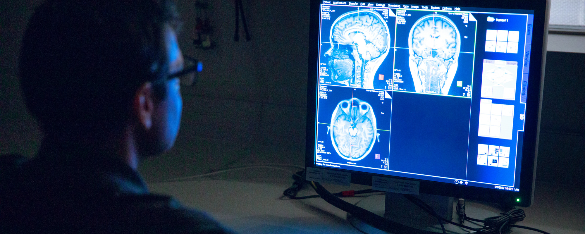
The MRI Research Facility is the centerpiece of UA’s growing focus on neuroscience research, allowing for the examination of the human brain and its development.

The facility has about 9,700 square feet of space comprising two MRI bays along with research, training, and administrative offices.

The facility is designed with space for behavioral data collection including biospecimen collection and processing, electroencephalography data collection and an MRI simulator.

The Alabama Life Research Institute oversees the facility.

Facilities & Equipment

Siemens 3T Prisma

This is a 60 cm bore whole-body MR system that is designed for high-performance human brain imaging. A host of coils are available including three head coils (64 channel head/neck, 32 channel, and 20 channel) and an assortment of coils for abdominal and orthopedic scanning. This scanner yields data with excellent signal to noise ratio, high stability, and excellent spatial resolution, and is capable of running Human Connectome Project complaint protocols, including high spatial and temporal resolution BOLD (fMRI) and diffusion-weighted imaging scans using multiband sequences. There is also a Nordic visual presentation display, an MR-compatible eye-tracking system, and behavioral response devices.

Magstim Geodesic EEG System

EGI’s proprietary Geodesic Sensor Net provides a simple method to apply dense arrays of sensors quickly and easily — you can apply up to 256 sensors in just a few minutes. The GSN gently holds each silver/silver chloride electrode sensor in place without the need for excessive head measurements or glues. And no scalp abrasion is necessary to get high-quality EEG data. Use with a simple saline solution for shorter recordings, or an inexpensive paste or gel for long-term recording.

MRI Mock Scanner

Coming soon!

Research Projects

Down Syndrome Study

Downs Syndrome Study: led by Fran Conners and Ian McDonough

HEALthy Brain and Child Development Study

UA, in partnership with UAB, is a data collection site for the HEALthy Brain and Child Development Study. The UA site is led by Drs. Lea Yerby and Sharlene Newman. UA Co-PIs include Joy Bradley, Holly Horan, John McDonald, Cathrine Lavendar and Brian Gannon. 

Resources

Prepping for a Scan

YOU CANNOT PARTICIPATE IF ANY OF THE FOLLOWING CONDITIONS APPLY:

  • If you have metal that is not MRI safe at 3T in your body from surgery, or have worked with metal that may have  resulted in metal shavings in your eye
  • If you are claustrophobic
  • For brain imaging, if you have permanent metal retainers or braces (fillings are fine)

When you come to scan, please

  • Wear comfortable clothing that do not contain metal (e.g., underwire bras, underwear with metallic threads including dry fit clothing)
  • Do not wear makeup with metal flakes including eye makeup
  • remove body piercings and jewelry 

Prior to scan, please make sure the researcher is aware of

  • the use of medication patches
  • if using an IUD (including the brand)
  • if you have any medical implants (e.g., screws or plates)
  • if you require glasses to correct vision (and you do not wear contacts); bring prescription
  • if you wear a medication patch (e.g., what is it, location)

Staff