Download equipment list as a Word document
Siemens Prisma 3T Scanner
The facility houses a 3T Siemens Magnetom Prisma scanner. The system has a gradient performance of 80 mT/m which allows for high resolution imaging with reduced acquisition times. The MRI system also includes the following coils used in brain, abdominal, and musculoskeletal imaging:
- 64-channel Head Coil
- 32-channel Head Coil
- 20-channel Head Coil
- Large and Small Body matrix coil
- Large and Small Flex coil
- Wrist Coil
- Foot/Ankle coil
- Knee Coil
- Large and Small Shoulder Coil
These coils, in conjunction with various pull sequence packages, make it a great resource for biomedical imaging research.
Magstim Geodesic EEG System
EGI’s proprietary Geodesic Sensor Net provides a simple method to apply dense arrays of sensors quickly and easily — you can apply up to 256 sensors in just a few minutes. The GSN gently holds each silver/silver chloride electrode sensor in place without the need for excessive head measurements or glues. And no scalp abrasion is necessary to get high-quality EEG data. Use with a simple saline solution for shorter recordings, or an inexpensive paste or gel for long-term recording.
MRI Mock Scanner
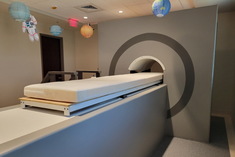
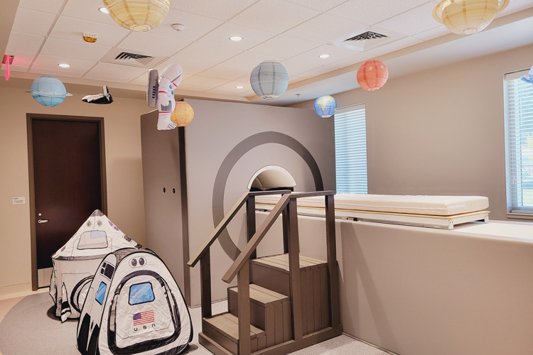
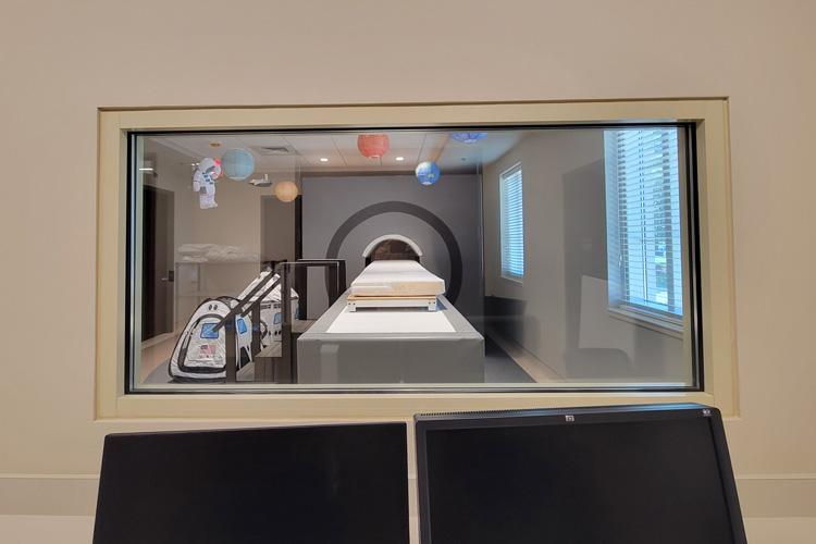
MRI mock scanner can provide a realistic MRI experience for those who feel anxious about being the actual scanner. The mock scanner can help determine whether a person can be comfortable being in a real MRI scanner and their ability to do the necessary tasks during a study. Mock scanner is equipped to play sounds typically heard during MRI scans. We have decorated the mock scanner space with a kid-friendly space exploration theme. This allows us to show how being in MRI is like being in a spaceship, with the MRI head coil being the helmet for their spacesuit.
We encourage research teams to arrange a visit to the mock scanner if any of your participants express anxiety about being in the scanner. This will give them an opportunity to see for themselves, if they can handle being in a MRI scanner, and in some cases get over their anxiety of being in small spaces.
The mock scanner is in the same facility as the Siemens Prisma.


Nordic Neurlabs: InRoom Viewing Monitor
Imaging studies that include visual stimuli can be displayed on a 40”, 4K (2160p) MRI compatible display. The display can be rolled behind the scanner bore and adjusted in way that allows participants to view the visual stimuli or instructions on that screen using a periscopic mirror. The monitor is shielded to prevent any interference with the MRI signal.
The monitor includes a camera on the top frame, which enables the researchers to monitor the participant during the scans. The screen can be flipped horizontally or vertically to display and image for participant lying on the scanner in any orientation. The monitor accepts inputs through variety of video adapters, which are then converted to optical signals that are sent over fiber optical cable through the waveguide to connect with the monitor to prevent any electromagnetic interference from entering the scanner room.
fORP package for responses recording
Some studies need input from participants in response to certain stimuli. These can be recorded using MRI compatible response button package that includes:
- FIU-932 response device
- 1 x 2 response button box
- 1 x 4 response button box
- 2 x 2 response button box
FIU-932 device connects to either the 1×2, 1×4 or 2×2 button box through a fiber optic connector which prevents them from causing interference inside the scanner room. Of these, the 1×2 and 1×4 response boxes allow a participant to response with one hand, either left or right. The 2×2 response box allows them to respond with 2 buttons on each hand.
The FIU-932 device connects to the presentation computer via USB and can be configured to send responses that are coded alphanumerically as required by the experimental design. This device can also read-in the synchronization pulses sent by the scanner at the onset of each acquisition or repetition time and transmit it to the presentation computer to use as time markers.
Opto-acoustic microphone: FOMRI-III + NC
Experiments that require the participant in the scanner to respond vocally can pose a unique challenge since the sound during BOLD scans can be very loud in comparison to human voice. To overcome that difficulty, OptoAcoustics FOMRI-III+NC system uses a head coil mounted microphone that uses special sound modeling to isolate the vocal response given by the participant from the scanner sound. The sound file can be recording in conjunction with synchronization pulses from the scanner which can be used in BOLD modelling during analysis.
BIOPAC MP160 + Electrodermal Activity acquisition system
Along with AcqKnowledge software, the BIOPAC system includes a fully automated electrodermal response scoring tool that locates skin conductance responses, visually identifies them in the record and measures them. It also automates event related potentials (ERP) analysis by locating the onset of the stimuli and identifying a valid SCR.
MRI compatible headphones: MR Confon
Presenting audio to the participant during an MRI experiment requires the sound levels to be audible over the scanner sounds. MR Confon uses piezoelectric headphones that are MRI compatible and can present the sound from MRI experiment clearly while isolating the scanner sound from the study participants. There is an additional feature where you can use a microphone connected to the MR Confon amplifier that streams the audio directly into the headphones as opposed to playing it in the scanner room allowing the researcher to provide clear instructions to the participants during a scan.
Fully Integrated Real time MRI Monitoring: FIRMM
Participant movement during the scans can make the data collected during the study difficult to analyze or in some cases, completely unusable. Having the ability to track the motion of your study participant can improve the data quality or provide a metric for how much of the collected data is within the acceptable motion threshold. In addition to providing a live display of participant motion during the scan, there are ways to have a biofeedback presented to the participant, in forms of a gauge, fixation cross, or a motion plot. This feedback encourages to participants to stay within the range by challenging them to remain still during the course of data collection. This system can be used for T1/T2 scans that have a built-in navigator, BOLD , ASL and diffusion scans.
High Performance Computing (HPC) Clusters
The Office of Information Technology (OIT) manages two HPC clusters: UAHPC and CHPC. UAHPC follows a “condo model”, in which individual researchers purchase nodes and share unused cycles with other researchers while retaining priority use on their purchased nodes. Currently, UAHPC has 1664 cores across 54 nodes providing over 56 TFLOPs theoretical sustained double precision performance. The cluster includes three GPU nodes and three high memory nodes. Users are limited to 1500 jobs within a 24-hour period on UAHPC. All UAHPC MXXX nodes are connected internally within their Dell M1000e chassis via InfiniBand FDR10 at a throughput of 40 Gbps. C6420 and R series nodes are directly connected to external InfiniBand. All the chassis are interconnected through a pair of external InfiniBand switches at 56 Gbps or 100 Gbps (2:1 oversubscribed). Storage is shared between nodes using NFS on IPoIB.
CHPC, a CC* grant awarded HPC, is available to all researchers on an equal basis. CHPC has 2112 cores across 34 nodes. In addition to 25 compute nodes, there are four 2 CPU AMD 7532 48 core 2 TB high memory nodes and five Dell PowerEdge R7525 GPU nodes each with one NVIDIA A100 GPU and room for two additional GPU. CHPC has 80 TFLOPS theoretical sustained performance. If used for machine learning applications, the cluster is capable of 3.2 PFLOPs tenser double-precision with sparsity. Twenty percent of unused cycles on CHPC are shared via Open Science Grid (OSG). The clusters attach independently to a shared Panasas Ultra storage array with 750TB of usable storage. All CHPC nodes are connected internally within their Dell chassis via InfiniBand FDR10 at a throughput of 40 Gbps. All nodes are directly connected to external InfiniBand. All the chassis are interconnected through a pair of external InfiniBand switches at 56 Gbps or 100 Gbps (2:1 oversubscribed). Storage is shared between nodes using NFS on IPoIB.
MRI Supplies
- 2 interview rooms for neuropsychological testing.
- Wellness room for accompanying family members or nursing mothers.
- Biospecimen room for acquiring samples of blood, saliva, etc. and the capability to centrifuge and store samples in ultra-low temperature refrigerator that can go to -87°C.